Figure 1.

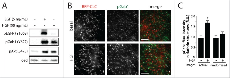
HGF stimulation increases phosphorylation of Gab1 and enrichment of pGab1 within CCPs. (A) RPE cells were stimulated with either 5 ng/mL EGF or 50 ng/mL HGF for 5 minutes. Whole-cell lysates were prepared, resolved by immunoblotting, and probed with anti-phospho-EGFR (Y1068), anti-phospho-Gab1 (Y627) and anti-phospho-Akt (S473) antibodies. (B-C) RPE cells stably expressing Tag-RFP-T fused to clathrin light chain (RFP-CLC) were stimulated with 50 ng/mL HGF and immunostained for pGab1. Shown in (B) are representative micrographs obtained by TIRF-M (scale 5 μm, arrowheads indicate pGab1-positive CCPs, selected manually). Images obtained by TIRF-M were subjected to automated detection of clathrin structures followed by quantification of pGab1 and RFP-CLC in each detected object, as in.5 Shown in (C) is the mean pGab1 fluorescence intensity detected within clathrin structures in the presence and absence of HGF compared to randomized control (> 35 cells per condition from 3 independent experiments).
