Figure 1.
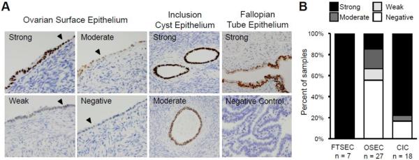
PAX8 expression in normal ovarian surface epithelium. (A) PAX8 expression was evaluated in 27 normal ovaries using immunohistochemistry. PAX8 expression ranged from strong to negative in OSECs and was strong, moderate or negative in CIC epithelial cells. FTSECs stained positive for PAX8. Examples of normal OSEC morphology on the ovarian surface are indicated with a black arrowhead. Tissue sections are shown at 100x magnification, negative control shown at 40x. (B) Graphical illustration of the range and proportion of tissues with PAX8 expression in primary OSEC, CIC, and FTSEC tissue. OSEC = ovarian surface epithelial cell, CIC = cortical inclusion cyst, FTSEC = fallopian tube secretory epithelial cell.

