
Figure 1. Ruxolitinib synergizes with MMF to kill brain, lung and triple negative breast cancer cells.
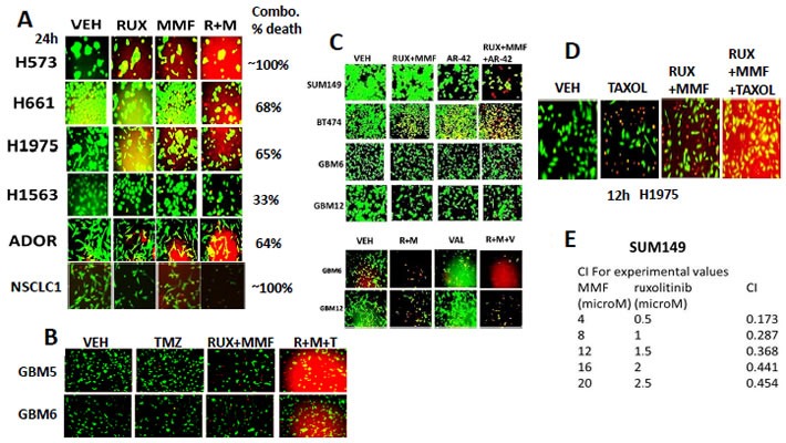
A. Non-small cell lung cancer cells were treated with vehicle control, ruxolitinib-phosphate (2.5 μM), MMF (5.0 μM) or the drugs in combination. Twenty four h later cell viability was assessed using a live/dead assay in a Hermes WiScan microscope at 10X magnification (n = 3 +/− SEM). B. GBM5 and GBM6 cells were treated with vehicle control, Temozolomide (TMZ, 50 nM), [ruxolitinib (1 μM) + MMF (5 μM)], or the three drugs in combination. Twelve hours later, cells were isolated and processed. Cell viability was assessed using a live/dead assay in a Hermes WiScan microscope at 10X magnification (n = 3 +/− SEM). C. Upper: GBM6, GBM12, SUM149 and BT474 cells GBM6, GBM12 and SUM149 cells were treated for 12h with vehicle control or with ruxolitinib (1.0 μM) and MMF (5.0 μM) in the presence of vehicle control or with the HDAC inhibitor AR-42 (0.3 μM). Twelve h after drug exposure cell viability was assessed using a live/dead assay in a Hermes WiScan microscope at 10X magnification. Lower; GBM6 and GBM12 cells were treated for 12h with vehicle control or with ruxolitinib (1.0 μM) and MMF (5.0 μM) in the presence of vehicle control or with the HDAC inhibitor Sodium valproate (0.75 μM). Twelve h after drug exposure cell viability was assessed using a live/dead assay in a Hermes WiScan microscope at 10X magnification. D. H1975 cells were treated with vehicle control, [ruxolitinib (1.0 μM) and MMF (5.0 μM), paclitaxel (10 nM) or the drugs in combination. Twelve h after drug exposure cell viability was assessed using a live/dead assay in a Hermes WiScan microscope at 10X magnification. E. SUM149 cells were plated (250-1,000) cells per well of a six well plate and 12h after plating were treated with vehicle, ruxolitinib (0.5-2.5 μM), MMF (4-20 μM) or in combination at a constant ratio for 24h, as indicated. The media was removed, cells were washed with drug free media, and the cells cultured for another 10 days in drug free media. Cell colonies were fixed, stained and groups of cells > 50 were counted as colonies. The combination index (CI) for synergy was calculated using the Calcusyn for Windows program using the Cho and Tallalay Method (n = 2; 12 individual wells per data point +/− SEM). A combination index of less than 0.70 indicates a strong level of tumor-killing synergy between the drugs.