
Figure 6. [Ruxolitinib + MMF] exposure increases ROS levels and causes release of AIF into the cytosol and AIF localization in the nucleus.
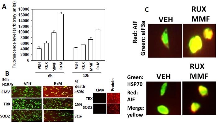
A. SUM149 cells were treated with vehicle control or with, ruxolitinib (1.0 μM), MMF (5.0 μM)] or in combination for 6h and 12h. Fifteen minutes before each time point cells are incubated with diacetate ester dichlorofluorescin (DCFH), 5 μM. The conversion of DCFH to DCF by reactive oxygen and nitrogen species is measured in sextuplicate in a Vector 3 plate reader (n = 3 +/− SEM).B. SUM149 cells were transfected with empty vector control (CMV) or with plasmids to express super-oxide dismutase 2 (SOD2) or thioredoxin (TRX). Twenty four h after transfection cells were treated with vehicle control or with, ruxolitinib (1.0 μM), MMF (5.0 μM)] or in combination for 36h. Cell viability was assessed using a live/dead assay in a Hermes WiScan microscope at 10X magnification (n = 3 +/− SEM). C. Upper: GBM12 cells were treated with vehicle control or with [ruxolitinib (1.0 μM) + MMF (5.0 μM)] for 6h. After 6h cells were fixed in place and permeabilized using 0.5% Triton X100. Immuno-fluorescence was performed to detect the expression of AIF (red fluorescent stain); eIF3A (green fluorescent stain) with images at 60X magnification. Lower: GBM12 cells were treated with vehicle control or with [ruxolitinib (1.0 μM) + MMF (5.0 μM)] for 6h. After 6h cells were fixed in place and permeabilized using 0.5% Triton X100. Immuno-fluorescence was performed to detect the expression of AIF (red fluorescent stain) and HSP70 (green fluorescent stain). The co-localization of AIF with HSP70 was determined by merging the images in Adobe Photoshop CS6 at 9999 dpi.