
Figure 3. Tau increases mitochondrial membrane potential and induces mitofusin accumulation.
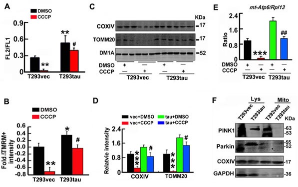
A. T293tau or T293vec cells were treated with CCCP (20 μM) or DMSO (vehicle control) for 30 min, then the membrane potential (FL2/FL1) was measured by JC-1 staining (n = 10). B.T293tau or T293vec cells were incubated with TMRM (20 nM) for 30 min, and then the fold ΔTMRM+ intensity (ΔΨm) was analyzed. C.-E. Levels of mitochondrial marker proteins (COX IV and TOMM20) and ratio of mt-Atp6/Rpl13 were detected by Western blotting (C, D) and real-time PCR (E), respectively, in T293tau and T293vec cells after treatment with CCCP (20 μM for 30 min). (F) Levels of PINK1 and Parkin in cell lysates (Lys) and mitochondria (Mito) fractions measured by Western blotting. Data were expressed as mean±SD. *, p < 0.05; **, p < 0.01, ***, p < 0.001 vs T293vec+DMSO; #, p < 0.05, ##, p < 0.01 vs T293tau+DMSO.