
Figure 5. A direct interaction of tau with mitochondrial marker proteins.
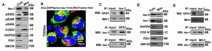
A. The cytoplasmic (Cyto) and mitochondrial (Mito) fractions were prepared from T293tau cells, and tau was analyzed by using a panel of phosphorylation site-specific antibodies, including tau-1 (reacts with the unphosphorylated tau at Ser198/199/202), pS262, pS396 and pS404. B. The representative image shows co-staining of tau-1 (green) with MitoTracker Red in T293tau cells. C. Co-immunoprecipitation data show association of tau with mitochondrial marker proteins OPA1 and Mfn1. D. The cytoplasmic (Cyto) and mitochondrial (Mito) fractions were prepared from the hippocampus of htau transgenic mice, and tau was analyzed by using anti-tau-1 antibody. E. Co-immunoprecipitation data show association of tau with mitochondrial marker proteins OPA1 and Mfn1 in hippocampal extracts of htau transgenic mice. GAPDH is a marker of cytoplasmic proteins, while TOMM40 and COXIV are respectively markers of mitochondrial outer membrane, inner membrane and intermembrane space proteins. PDI or GM130 is the marker of ER or Golgi apparatus used to prove the purity of the mitochondrial fraction.