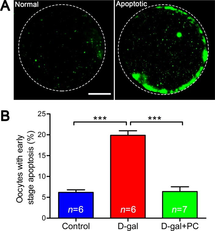
Figure 8. PC inhibited early stage apoptosis in MII oocytes in D-gal-induced aging mice.
A. Representative images of early stage apoptosis in MII oocytes. Oocytes without green fluorescence signals at the zona pellucida and oocyte membrane were non-apoptotic, and oocytes undergoing early apoptosis were characterized by a clear green signal in the zona pellucida and membrane. Scale bar = 20 μm. B. Percent oocytes undergoing early stage apoptosis. D-gal induced early stage apoptosis in oocytes, and this was inhibited by PC. Data are presented as the means ± SEMs and were processed by one-way ANOVA and Newman-Keuls post hoc tests. Significant differences between groups, ***P < 0.001. n indicates the number of mice for each treatment.

