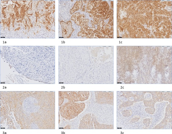
Figure 2. Examples of (dis)concordance in FRα staining in biopsy-, primary tumor-, and metastatic LN tissue in NSCLC and breast cancer patients.

1a-1c: example of concordance between positive FRα expression on biopsy (1a), primary tumor (1b) and metastatic LN tissue (1c) in a NSCLC patient, containing adenocarcinoma (20x). 2a-2c: example of disconcordance between FRα expression on biopsy (2a), primary tumor (2b) and metastatic LN tissue (2c) in a NSCLC patient, containing SCC (20x). 3a-3c: example of concordance between positive FRα expression on biopsy (3a), primary tumor (3b) and metastatic LN tissue (3c) in a breast cancer patient (20x).
