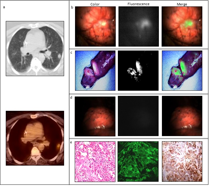
Figure 4. Example of FRα-targeted detection of an adenocarcinoma using EC-17, i.e.

fluorescent FRα-targeted molecular agent, in a patient suffering from NSCLC. In vivo fluorescence imaging was performed using the Artemis imaging system [34]. a: Prior to pulmonary resection, a tumor of 3 cm in the upper lobe of the left lung is detected by CT-and PET-scan. b: In vivo fluorescence imaging shows clear tumor delineation. c: Ex vivo fluorescence imaging of the tumor in the resected specimen. d: After resection, the wound bed was inspected with λex 490 nm and demonstrated no residual fluorescence at the surgical margins. e: FRα upregulation was confirmed by fluorescence microscopy and immunohistochemical staining.
