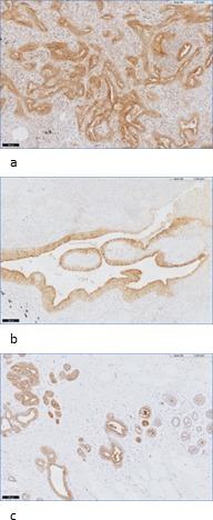
Figure 5. Examples of FRα staining in normal lung and breast tissue.

a: staining of FRα at the luminal border of normal lung tissue in an adenocarcinoma patient (10x). b: staining of FRα at the luminal border of normal lung tissue in a SCC patient (10x). c: staining of FRα in normal breast tissue: staining at the luminal border of secretory cells (10x).
