
Figure 1. FoxO1 is increased in alternately activated alveolar macrophages.
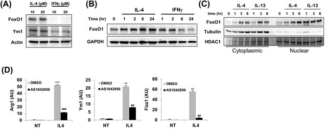
A. FoxO1 expression in different subsets of MH-S alveolar macrophages. MH-S cells were stimulated with IL-4 or IFN-γ for 24 h. FoxO1 is virtually eliminated in cells by IFN-γ. B. Immunoblot of FoxO1 in control or IL-4 or IFN-γ-treated macrophages for 0-24 h. C. Cytoplasmic and nuclear extracts were prepared from IL-4 and IL-13-stimulated MH-S alveolar macrophages, and FoxO1 localization was measured by Western blot analysis. D. qPCR analysis of Arg1, Ym1, Fizz1 mRNA expression by MH-S pretreated with FoxO1 inhibitor AS1842856 (1μM) and stimulated with for 24 h with IL-4 (10 ng/ml). Data are representative of at least three independent experiments (A-D). **p < 0.01, ***p < 0.001 vs. nontreated, ##p < 0.01, ###p < 0.001 vs. IL-4 with noninhibitor (student's t-test).