
Figure 4. LysMFoxO1Tg mice showed impaired development of DRA-induced allergy airway inflammation.
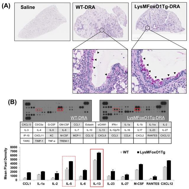
A. Histopathology was performed based on H&E staining to determine the asthmatic inflammation in Saline- or DRA-treated WT or LysMFoxO1Tg mice. Upper panel shows H&E staining for the entire left lungs. Lower panel shows a zoomed section of the lung as indicated by a square in the upper panel. PAS-stained lung sections from the mice exposed to PBS or DRA. Black arrowheads indicate PAS-positive goblet cells. B. Cytokines were detected in BAL fluid from DRA-challenged WT (LysM) and LysMFoxO1Tg mice was quantified and normalized to WT using an antibody array.