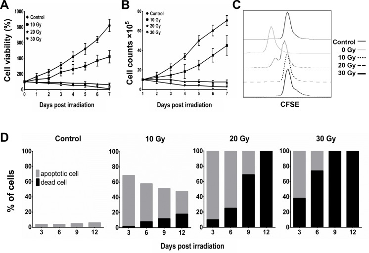
Figure 3. Inhibition of proliferation and induction of apoptosis of BA15 by γ-ray irradiation.
After different dosages of irradiation, the cell viability and proliferation of BA15 cells were analyzed by MTT, cell counting, and CFSE assays. Apoptosis assays were performed every 3 days after irradiation. A. MTT assay indicated that the cell viability of BA15 cells decreased after exposure to 20 Gy and 30 Gy of irradiation. B. Cell counting indicated that the number of BA15 cells decreased after exposure to 20 Gy and 30 Gy of irradiation. C. CFSE labeling revealed that the proliferation of BA15 cells was completely inhibited after exposure to irradiation of 20 Gy and 30 Gy. D. The apoptosis assay revealed that all the cells in the 20-Gy and 30-Gy group were in apoptosis or dead 3 days after irradiation and that all the cells had died by day 12. There were fewer dead cells in the 20-Gy group than in the 30-Gy group at every time point. Error bars indicate standard deviations.

