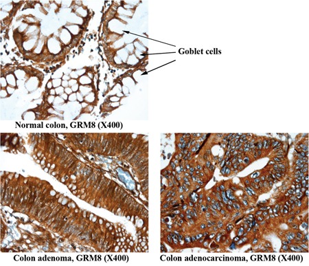
Figure 3. Representative images of non-neoplastic normal colon, colon adenoma and colon adenocarcinoma tissue staining of GRM8.

Arrow indicates goblet cells containing mucin in normal colon epithelium. The mucin is digested and lost during the immunohistochemical procedure, leaving the cytoplasm empty and negative for the marker assessed. The conventional colonic adenocarcinoma cells produce less mucin and exhibit a higher IHC score.
