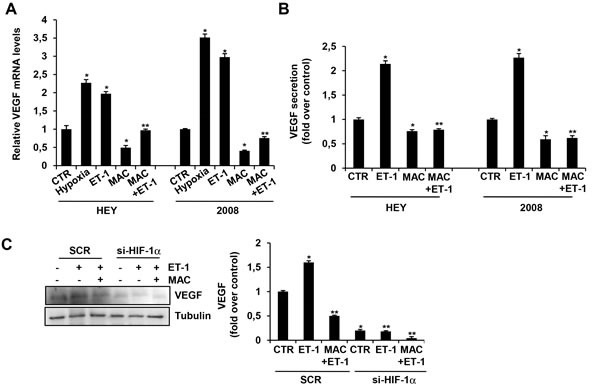
Figure 1. HIF-1α mediates ET-1/ETAR axis-induced VEGF expression that is inhibited by macitentan in EOC cells.

A. HEY and 2008 EOC cells were cultured in serum free-media under normoxic or hypoxic conditions for 24 h or treated with ET-1 in the absence or in the presence of macitentan (MAC), and VEGF mRNA expression was analyzed by qPCR. Bars are means ±SD from three independent experiments each performed in triplicate. *, p < 0.01 versus CTR; **, p < 0.01 versus ET-1. B. HEY and 2008 cells were stimulated with ET-1 in the absence or in the presence of MAC. VEGF protein secretion was analyzed by ELISA in EOC cell conditioned media collected after 24 h. Bars are means ±SD from three independent experiments each performed in triplicate. *, p < 0.01 versus CTR; **, p < 0.001 versus ET-1. C. HEY cells were transfected with SCR or siRNA against HIF-1α and stimulated with ET-1 in the absence or in the presence of MAC. VEGF protein levels were analyzed by immunoblotting (IB). Tubulin was used as loading control. Densitometric analysis (right panel) of VEGF protein bands from three independent experiments, normalized to tubulin content. Bars are means ±SD. *, p < 0.01 versus CTR; **, p < 0.001 versus ET-1.
