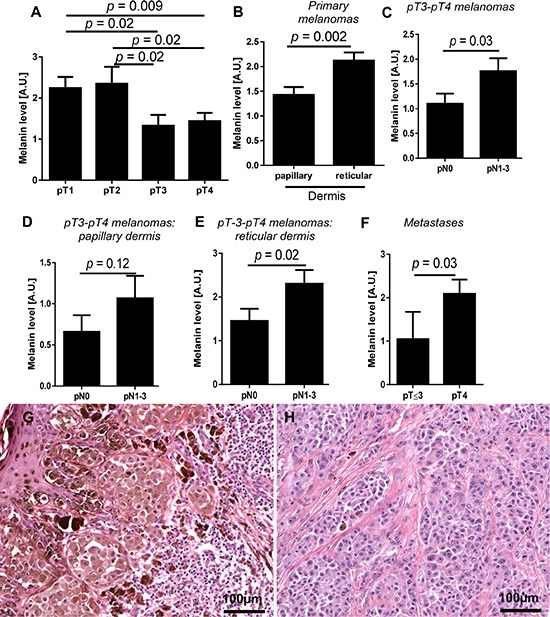
Figure 2.

Mean melanin level in primary melanomas in relation to pT status (A) n = 84), localization of melanoma cells in the skin (B) n = 84) and pN status in pT3-pT4 melanomas (C). Melanin level in melanoma cells localized within papillary (D) and reticular dermis (E) of non-metastasizing (pN0) and metastasizing (pN1-3) pT3-4 melanomas (n = 45). Melanin level in metastases developed in pT2-3 and pT4 melanomas (F). Representative pT1 (G) and pT4 (H) melanoma cases. The p values represent statistical significance in Mann-Whitney test.
