
Figure 2. WDR26 is overexpressed in cell lines and tissue samples of human breast cancer.
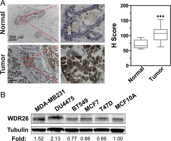
A. WDR26 expression is significantly increased in malignant human breast cancer samples, as compared to normal breast tissues. Representative images show immunohistochemical staining of WDR26 protein in human breast cancer samples (Stage III) and matched, adjacent, normal tissues. Quantitative data are shown on the right. *** p<0.001 (n=15). B. Western blot analysis of WDR26 expression in human breast cancer cell lines. The relative level of WDR26 expression is expressed as a fold change over that in MCF10A (after normalization by tubulin) and indicated underneath the blot (n=3).