
Figure 1. Qualification of DRG and staining of TRPV1, SP, and CGRP in DRG neurons.
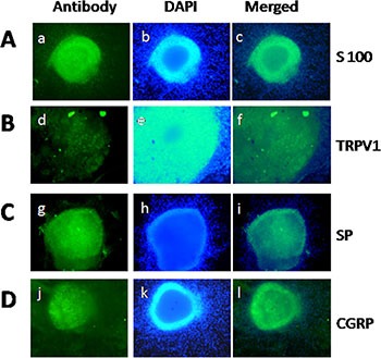
(A) Identification of DRG extracted from newborn rat by S100 staining. (B–D) Staining of TRPV1, SP, and CGRP in DRG neurons. (a, d, g, j) S100, TRPV1, SP, and CGRP-positive neurons (green) in DRG, respectively. (b, e, h, k) Nuclear staining with DAPI. (c, f, i, l) Merge of staining is shown.