
Figure 1. Expression of L-FABP and VEGF-A in tissues obtained from HCC patients.
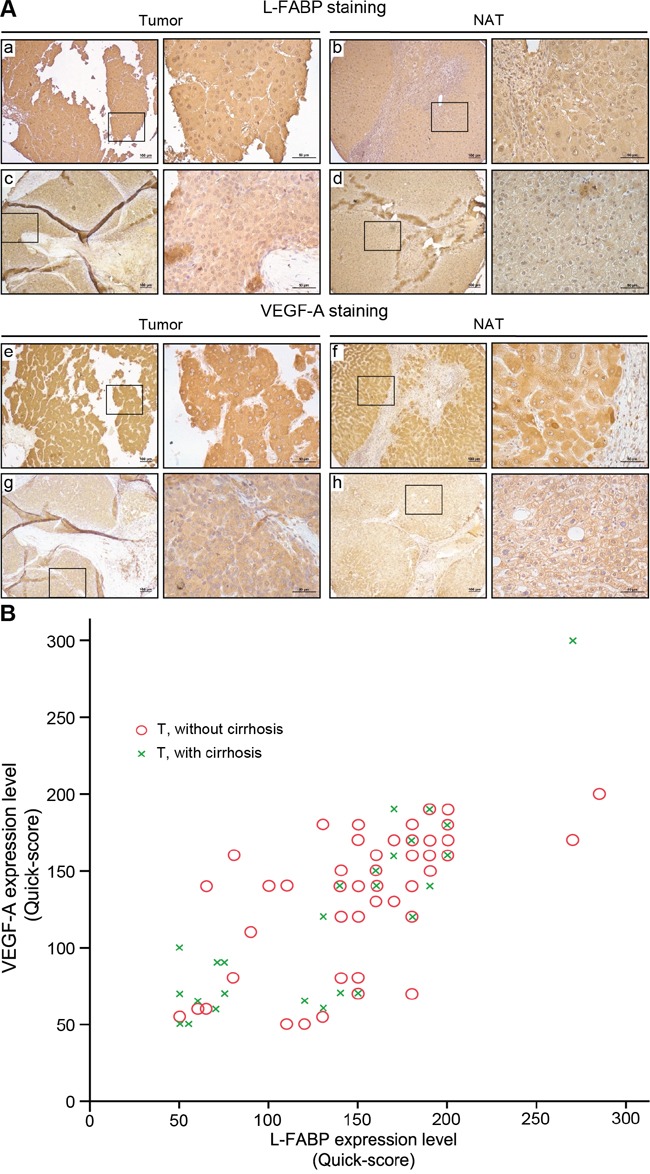
Protein expression was assessed in 90 HCC cases using immunohistochemical staining of paired normal (NAT) and tumor tissues. A. Staining of L-FABP and VEGF-A was observed in tumor tissues (L-FABP: a and c; VEGF-A: e and g) and their paired normal adjacent tissues (L-FABP: b and d; VEGF-A: f and h). Staining intensity: a and e, strong; b, c, f, and g, moderate; d and h, weak. B. Positive correlation between L-FABP and VEGF-A expression in 90 HCC tissues with and without cirrhosis (Pearson's correlation coefficient, r = 0.737; p < 0.01).