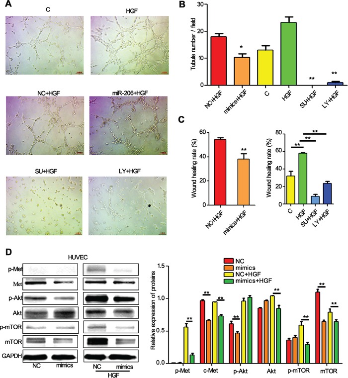
Figure 7. miR-206 suppresses the migration and tube formation of HUVEC cells.

A, B. Representation (A) and quantification (B) of tube formation assay showing the angiogenic capability of HUVECs transfected with NC or miR-206, and then stimulated with HGF (50 ng/ml). Original magnification, ×100. * P<0.05 vs NC+HGF, ** P<0.01 vs HGF C. Quantification of scratch migration assay showing the migration of HUVEC cells transfected with NC or miR-206, and then stimulated with HGF (50 ng/ml). D. HUVECs transfected with NC or miR-206 were stimulated with 20 ng/ml of HGF for 15min. The cells were then harvested and lysed for the detection of p-c-Met, c-Met, p-Akt, Akt, p-mTOR, mTOR, and GAPDH. C: control; NC: Negative control; SU: SU11274; LY: LY294002 ** P<0.01.
