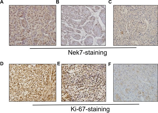
Figure 2. Immunohistochemistry of Nek7 and Ki-67 on HCC specimens.

Representative immunohistochemical staining of HCC specimens with different histologic grade, as determined using anti-Nek7 and anti-Ki-67 antibodies. A. The expression status of Nek7 in low-grade HCC tissue. B. An example of moderate-differentiated HCC with Nek7 expression. C. Nek7 expression in high-grade HCC tissue. D. Ki-67 expression status in low-grade HCC tissue. E. Example of moderate-differentiated HCC with Ki-67 expression. F. Ki-67 expression in high-grade HCC tissue.
