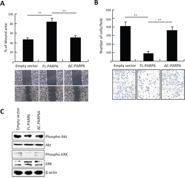
Figure 2. PARP6 inhibits invasion and migration.

A. Empty vector, FL-PARP6 and ΔC-PARP6 transfectant cells were subjected to wound healing assays. The uncovered areas as a percentage of the original wound area were quantified. **, P < 0.01 is based on the Student t test. All results were from three independent experiments. Error bars, SD. B. Empty vector, FL-PARP6 and ΔC-PARP6 transfectant cells were subjected to in vitro invasion assays using matrigel. Number of cells that invaded through matrigel was counted. We performed the assay 3 times and 5 randomly selected fields from each membrane were counted under a light microscope at x100 magnification. **, P< 0.01 is based on the Student t test. Error bars, SD. C. Expression of phospho- and total protein of Akt and ERK in empty vector, FL-PARP6 and ΔC-PARP6 transfectant SW480 cells by Western blot analysis. β-actin expression was used as a loading control.
