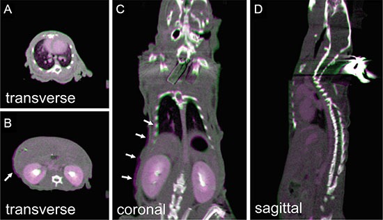
Figure 6. The pseudocolor overlay of mouse CBCTs acquired immediately (purple) and 15 minutes (green) after animal setup on the stage.

The animal received iodine contrast injection before imaging. (A–D) are one transverse slice at thorax region, transverse slice at abdominal region, coronal slice, and sagittal slice, respectively. Arrow in (B and C) point to the slight mismatch at the ribs and abdominal skin contours.
