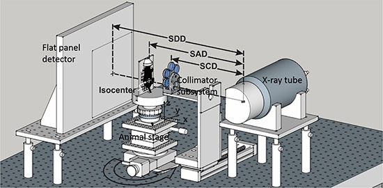
Figure 7. The iSMAART configuration.

The x-ray tube and flat-panel detector are fixed on the optical bench and the animal stage can be translated along x-y-z directions and rotated around z-axis. The anesthetized mouse is positioned upright with a customized animal holder. The motorized collimation subsystem is positioned in or out of the radiation for treatment or imaging. The imaging and irradiation isocenters are coincident with each other. The nominal values for the source to detector distance (SDD) is 52.5 cm, source to axis distance (SAD) 35 cm, and source to collimator distance (SCD) 25 cm.
