
Figure 5.
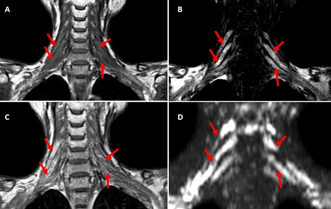
A-D. The coronal MRI scan of one case with bilateral RIBP T1-weighted coronal MRI scan showed bilateral brachial plexus were swollen with hyper-intensity. (A) C-5∼C-8 roots and corresponding trunks of brachial plexus showed prolonged T2 relaxation time at T2-weighted MR imaging (B), high intensity at Post-contrast T1-weighted imaging (C) and DWIBS (D) (indicated with red arrows).