
Figure 2. MiR-1 and -133b bind to PTBP1.
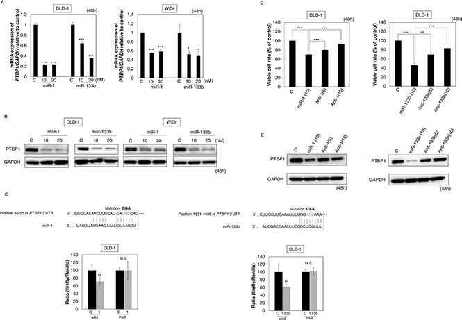
(A, B) The mRNA expression (A) and protein expression (B) of PTBP1 at 48 h after the transfection with miR-1 or -133b (10, 20 nM). (C) Luciferase activities after co-transfection with control, miR-1 or -133b and wild-type or mutant-type pMIR vectors having the predicted miR-1 or -133b binding site in the 3′UTR of PTBP1. The upper panel shows the region of the 3′-UTR of human PTBP1 mRNA complementary to the mature miR-1 or -133b. The box indicates the predicted binding sites for miR-1 or -133b. (D) Effect of combined treatment with antagomiR-1 / miR-1 or antagomiR-133b / miR-133b on the growth of DLD-1 cells. Cells were transfected with non-specific control, miR-1 or -133b (10 nM), miR-1 or -133b (10 nM) + antagomiR-1 or -133b (5 nM) or miR-1 or -133b (10 nM) + antagomiR-1 or -133b (10 nM). (E) Expression level of PTBP1 in DLD-1 cells assessed at 48 h after transfection of the cells. Results are presented as the mean ± SD; *P < 0.05; **P <0.01; ***P < 0.001; N.S., not statistically significant.