
Figure 8.
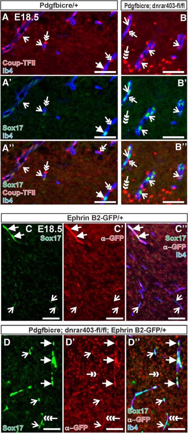
Elevated Sox17 expression in PdgfbiCre;dnRAR403-fl/fl venous and arterial vessels. A, B, Immunostaining for Sox17 (green) and Coup-TFII (red) on E18.5 PdgfbiCre/+ (A) and PdgfbiCre;dnRAR403-fl/fl (B) brains. Open arrows indicate Ib4+ (blue) vessels with Coup-TFII+ ECs (presumptive venous blood vessel). Arrow in A indicates blood vessel in control brain tissue that is Coup-TFII−/Sox17+ (presumptive arterial vessel). Double arrows indicate Coup-TFII+ mural cells; triple arrow indicates Coup-TFII+ neural cell. C, D, GFP (red) and Sox17 (green) immunostaining in control and PdgfbiCre;dnRAR403-fl/fl animals expressing Ephrin B2-GFP that labels arterial EC nuclei. Arrows indicate GFP+/Sox17+ arterial EC nuclei; open arrows indicate Sox17 expression in GFP- EC nuclei. C′′ and D′′ show overlay with Ib4 to label the vasculature (blue). Ephrin-B2-GFP is also expressed by some neurons (double-headed arrow). GFP IF is present in the endothelial cell membrane of images in D due to the IRES-GFP present in the PdgfbiCre allele construct (triple arrow). Scale bars, 100 μm.