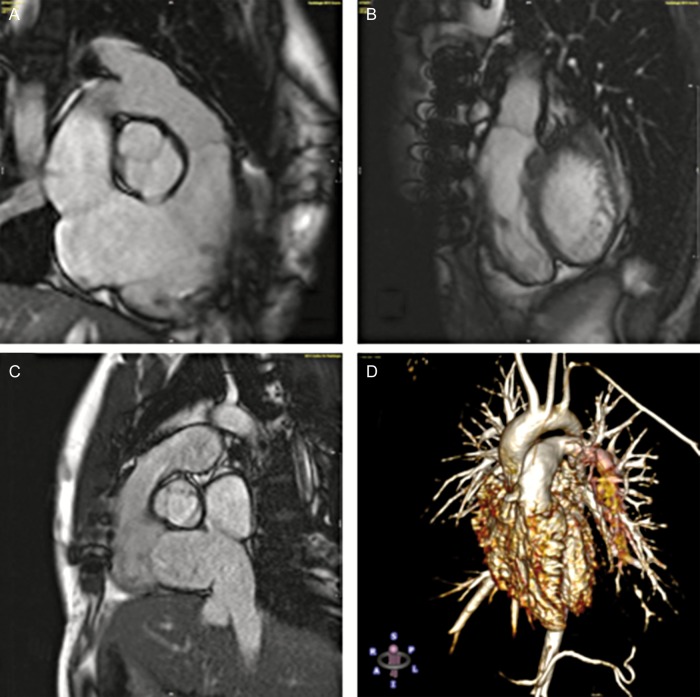
Figure 3:
Cardiac magnetic resonance imaging examples of DPH. (A) Coronary three-chamber view at diastole 72 months after DPH implantation in a 20-year old patient; (B) sagittal view of the patient (A) at diastole; (C) sagittal view of DPH at systole 78 months after implantation in a 24-year old patient; (D) contrast-enhanced angiography 116 months after DPH implantation in a 10-year old patient.

