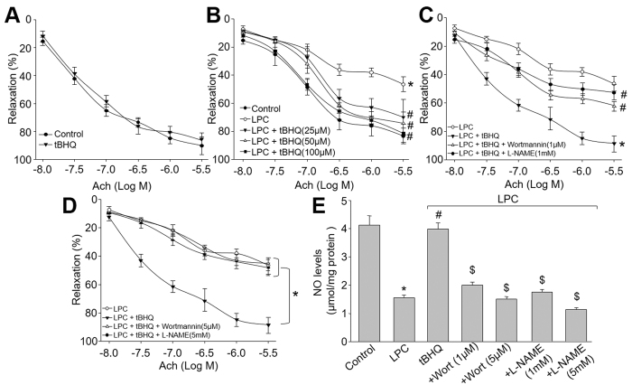
Figure 5. Inhibition of Akt or eNOS by pharmacological reagents abolishes tBHQ-prevented endothelial dysfunction ex vivo.
(A) The isolated mouse aortic rings were incubated with tBHQ (50 μM) for 12 hours and Ach-induced endothelium-dependent relaxation was assayed by organ chamber. (B) The isolated mouse aortic rings were preincubated with tBHQ (25, 50, 100 μM) for 30 minutes and then exposed to LPC (4 mg/l) for 2 hours. Ach-induced endothelium-dependent relaxation was assayed by organ chamber. *P < 0.05 vs. control, #P < 0.05 vs. LPC. (C) Isolated mouse aortic rings were preincubated with tBHQ (50 μM) for 30 minutes with or without wortmannin (1 μM) and L-NAME (1 mM) followed by LPC (4 mg/l) for 2 hours. The endothelium-dependent relaxation induced by acetylcholine was assayed by organ chamber. *P < 0.05 vs. LPC, #P < 0.05 vs. tBHQ. (D) Isolated mouse aortic rings were preincubated with tBHQ (50 μM) for 30 minutes with or without wortmannin (5 μM) and L-NAME (5 mM) followed by LPC (4 mg/l) for 2 hours. The endothelium-dependent relaxation induced by acetylcholine was assayed by organ chamber. *P < 0.05 vs. LPC, #P < 0.05 vs. tBHQ. (E) Homogenates of aortic tissues were subjected to measure NO productions by the Griess method in each group. *P < 0.05 vs. Control group. #P < 0.05 vs. LPC. $P < 0.05 vs. LPC + tBHQ.

