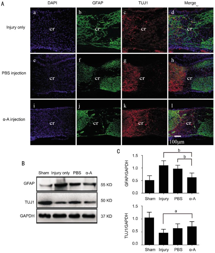
Figure 3. Intravitreous injection of αA-crystallin increased the number of TUJ1-positive processes at 14d after ONC.
A: Immunofluorescence staining showed the GFAP-positive fibers (green) and TUJ1-positive processes (red) in the crush site following ONC. The sections were obtained from the ONC injury group (injury only) (a-d), PBS-treated ONC injury group (PBS injection) (e-h) and αA-crystallin-treated ONC injury group (α-A injection) (i-l). B, C: WB analysis showed that the GFAP levels in the optic nerve were significantly decreased in the αA-crystallin-treated group compared to the injury only (P<0.01) and PBS-treated groups (P<0.01). However, the TUJ1 levels were higher in the αA-crystallin-treated group compared to the injury only group (P<0.05). cr: The middle of crush site; ONC: Optic nerve crush. The quantitative data represent the means±SD (n=5). aP<0.05, bP<0.01. Scale bar=100 µm.

