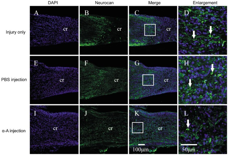
Figure 7. Intravitreous injection of αA-crystallin decreased the expression of neurocan in the crush site at 14d after ONC.
Immunofluorescence staining showed the neurocan (green) expression in the crush site following ONC in the ONC injury (injury only) (A-C, and C zoomed image D), PBS-treated ONC injury (PBS injection) (E-G, and G zoomed image H) and αA-crystallin-treated ONC injury groups (α-A injection) (I-K, and K zoomed image L). The arrows indicate neurocan-positive labeling. cr: The middle of crush site. The arrows indicate neurocan-positive labeling.

