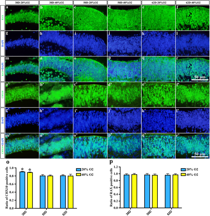
Figure 3. Neuroectodermal epithelium expressed the neural retina marker RAX and the retinal progenitor marker CHX10.
(a–f) RAX-immunoreactive neural retina tissue of embryonic bodies in (a) 38D 20% O2; (b) 38D 40% O2; (c) 50D 20% O2; (d) 50D 40% O2; (e) 62D 20% O2; (f) 62D 40% O2. (g–l) Corresponding DAPI staining in (a–f). (m–r) Merged image of RAX and DAPI. (a’–f’) CHX10-immunoreactive neural retina tissue of embryonic bodies in (a’) 38D 20% O2; (b’) 38D 40% O2; (c’) 50D 20% O2; (d’) 50D 40% O2; (e’) 62D 20% O2; (f’) 62D 40% O2. (g’–l’) Corresponding DAPI staining in (a’–f’). (m’–r’) Merged image of CHX10 and DAPI. (o) statisitical analysis of CHX10 positive cells. Asterisk showed the significant difference between different time point under same oxygen concentration. (p) Statisitical analysis of RAX positive cells.

