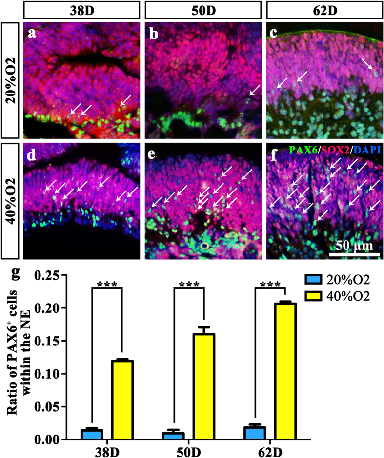
Figure 5. High oxygen concentration facilitates neural retinal development similar to the status in vivo.
(a–f) SOX2 and PAX6 double-staining of NE in (a) 38D 20% O2; (b) 50D 20% O2; (c) 62D 20% O2; (d) 32D 40% O2; (e) 52D 20% O2; (f) 62D 40% O2. White arrows point at the PAX6-immunoreactive cells within the NE. (g) Ratio of PAX6-immunoreactive cells within the NE in 6 groups. The number of PAX6 positive cells within the NE in the high oxygen groups was much higher compared to the normal oxygen concentration groups. NE: Neuroectodermal epithelium.

