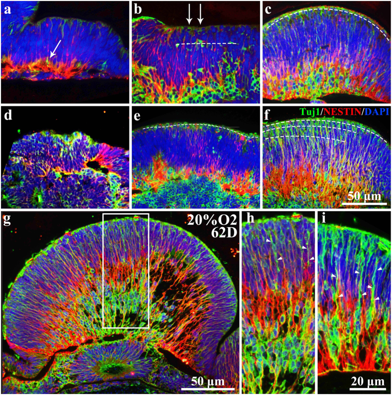
Figure 8. High oxygen concentration facilitated the proliferation, maturation and migration of RGCs in neuroectodermal epithelia of embryonic bodies.
(a–f) Tuj1 and NESTIN double-stained NE in (a) 38D 20% O2; (b) 50D 20% O2; (c) 62D 20% O2; (d) 32D 40% O2; (e) 52D 20% O2; (f) 62D 40% O2. White arrows in (a,b) point at the Tuj1-positive mature neurons. The dotted lines in (b, c, e, f) delineate the distribution of Tuj1-positive mature neurons. (g) Whole Tuj1/NESTIN double-stained NE picture in 62D 20% O2 group. (h) Enlarged images in (g). (i) Regional image of Tuj1/NESTIN double-stained NE in 62D 40% O2 group. White arrows in (h, i) point at the process of mature neurons. NE: Neuroectodermal epithelium; RGCs: retinal ganglion cells.

