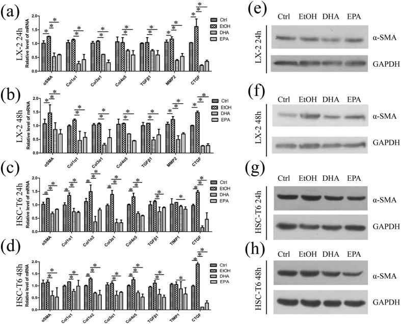
Figure 3. DHA and EPA down-regulate pro-fibrogenic genes in LX-2 and HSC-T6 cells.
(a–d) LX-2 (a,b) and HSC-T6 (c,d) cells were exposed for 24 h or 48 h to media containing 10% fetal bovine serum, ethanol, 75 μM DHA or EPA and then total RNA was isolated. The expression of α-SMA, collagen1α1, collagen1α2, collagen3α1, collagen4α5, mmp2, tgfβ1, timp1 and ctgf were measured by qPCR. (e–h) With the same treatment in (a–d) total protein of LX-2 (e,f) and HSC-T6 (g,h) cells were isolated and α-SMA was measured and quantified by western blot. GAPDH served as the loading control. The gels were cropped and the full-length gels are presented in Supplementary Fig. 5. The data are expressed as the mean ± SEM for triplicate experiments. *P < 0.05.

