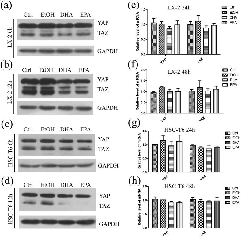
Figure 6. DHA and EPA decrease the protein level but not the mRNA level of YAP/TAZ in LX-2 and HSC-T6 cells.
(a–d) LX-2 (a,b) and HSC-T6 (c,d) cells were exposed for 6 or 12 h to media containing 10% fetal bovine serum, ethanol, 75 μM DHA or EPA. Cell lysates from both cell lines were subsequently assessed by immunoblot to determine the protein levels of YAP/TAZ. GAPDH was used as internal control. The gels were cropped and the full-length gels are presented in Supplementary Fig. 6. (e–h) The total RNA were extracted from LX-2 (e,f) and HSC-T6 (g,h) cells exposed for 24 h or 48 h to media containing 10% fetal bovine serum, ethanol, 75 μM DHA or EPA. The extracted RNA was subsequently used for qPCR. The data are expressed as the mean ± SEM for triplicate experiments. *P < 0.05.

