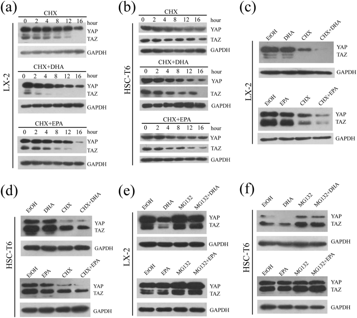
Figure 8. DHA and EPA induce YAP/TAZ degradation in LX-2 cells through proteasome-dependent pathway.
(a,b) LX-2 (a) and HSC-T6 (b) cells cultured in medium containing 10% FBS were treated with CHX alone, CHX in combination with 75 μM either DHA or EPA for the indicated durations. Cell lysates were prepared and used for immunoblot and GAPDH was used as a loading control. (c,d) LX-2 (c) and HSC-T6 (d) cells cultured in medium containing 10% FBS were treated with ethanol, 75 μM DHA or EPA, CHX, CHX in combination with 75 μM DHA or EPA for 12 h. Cell lysates were prepared and used for immunoblot and GAPDH was used as a loading control. (e,f) LX-2 (e) and HSC-T6 (f) cells were treated with ethanol, 75 μM DHA or EPA, MG132, MG132 in combination with 75 μM DHA or EPA for 24 h and were subsequently lysed for immunoblot. The gels were cropped and the full-length gels are presented in Supplementary Fig. 7. The data are expressed as the mean ± SEM for triplicate experiments. *P < 0.05.

