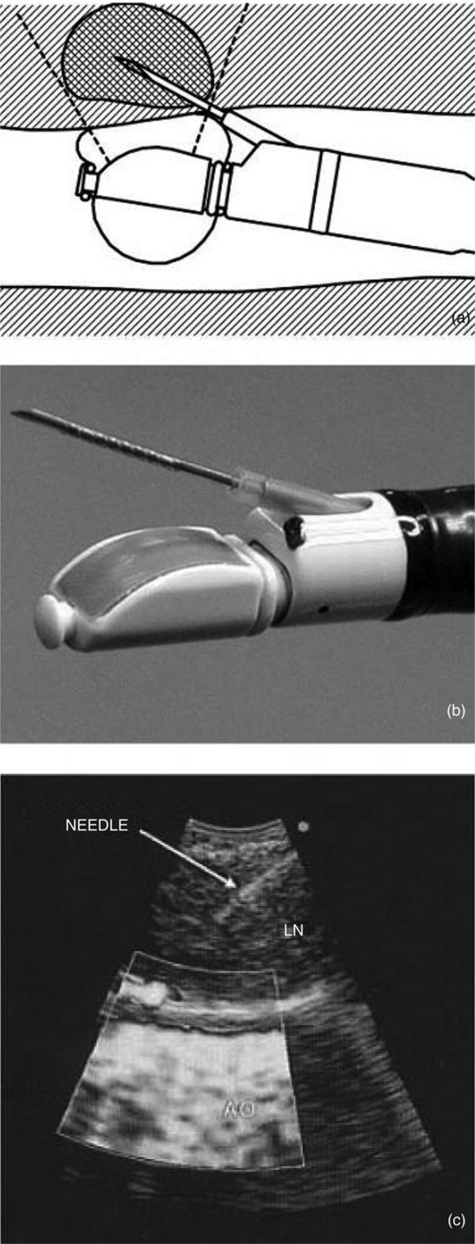
Fig 3.

a and b: (Linear probe) endobronchial ultrasound (EBUS)-guided transbronchial needle aspiration has the ultrasound transducer at the distal end of the EBUS bronchoscope. The direct view is 30° to the horizontal. The biopsy needle is placed through the working channel, extending from the end of the bronchoscope at 20° to the direct view. c: The linear ultrasound image (needle in a node) is a 50° slice, in parallel to the long axis of the EBUS bronchoscope (power Doppler flow image shown in bottom half). AO = aorta; LN = lymph node. Reproduced with permission of American College of Chest Physicians.5
