Figure 12.

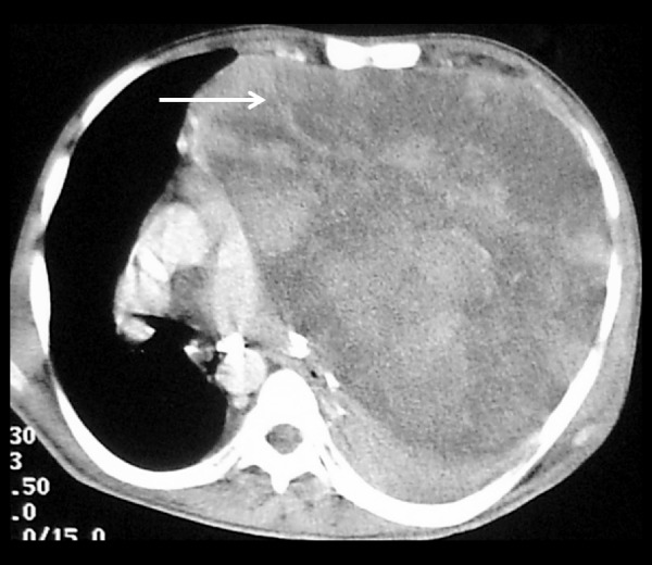
Liposarcoma: Axial CT section shows a large, heterogeneously-enhancing, necrotic mass occupying RSS and most of the left hemithorax (arrow). This was a biopsy-proven case of proven liposarcoma.

Liposarcoma: Axial CT section shows a large, heterogeneously-enhancing, necrotic mass occupying RSS and most of the left hemithorax (arrow). This was a biopsy-proven case of proven liposarcoma.