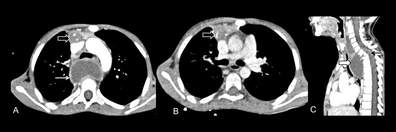
Figure 14.
(A, B) Tuberculosis: Axial CT sections show necrotic conglomerate nodes with abscess formation in the retrosternal region (black block arrow) along with a paravertebral abscess (white arrow). (C) Sagittal reformatted image shows Pott’s spine with paravertebral abscess (white block arrow) with epidural extension (black arrow).

