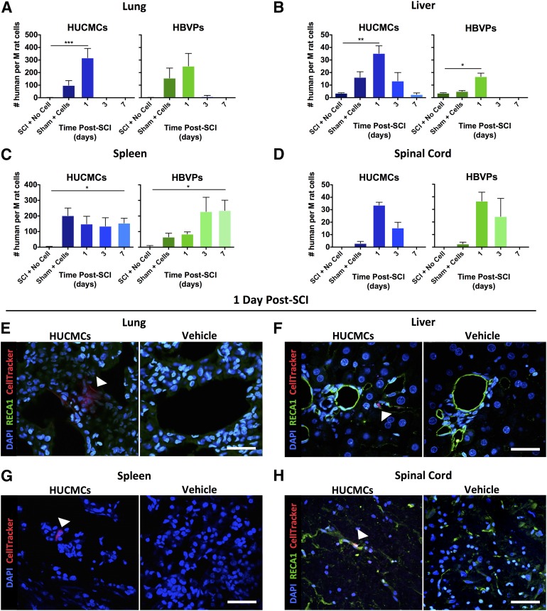
Figure 2.
Temporal human stromal cell bio-distribution after intravenous delivery following SCI reveals rapid cell clearance. (A–D): Quantitative reverse-transcription polymerase chain reaction for a human-specific sequence of DNA (thymidine kinase) was used to assess temporal cell (HUCMC and HBVP) clearance in the lungs (A), liver (B), spleen (C), and spinal cord (D). Data were converted to number of human cells (per million rat cells) and are expressed as mean ± SEM. (E–H): HUCMCs were also labeled with CellTracker dye before infusion for confocal imaging (×60, Nikon C2+ microscope) of their distribution in the lungs (E), liver (F), spleen (G), and spinal cord white matter (H) at 1 day (24 hours) after SCI. Arrows indicate CellTracker signal, One-way analysis of variance (Dunnett's multiple comparison). ∗, p ≤ .05; ∗∗, p ≤ .01; ∗∗∗, p ≤ .001; ∗∗∗∗, p ≤ .0001. Scale bar = 50 μm. Abbreviations: DAPI, 4′,6-diamidino-2-phenylindole; HBVPs, human brain vascular pericytes; HUCMCs, human umbilical cord matrix cells; M, million; RECA1, rat endothelial cell antigen 1; SCI, spinal cord injury.

