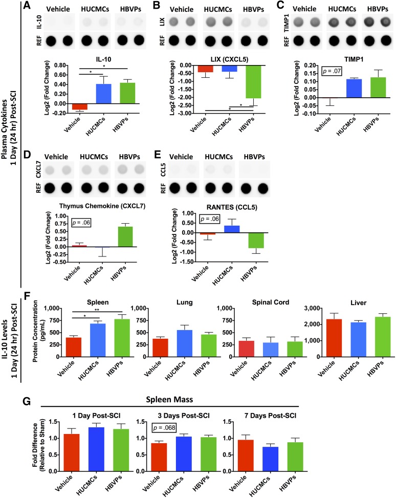
Figure 3.
Stromal cell infusion raises rodent circulating and splenic IL-10 levels at 1 day (24 hours) after SCI. Because very few cells were found in the spinal cord after intravenous infusion, we sought to characterize the systemic response. Rodent plasma cytokines changes were assessed with the R&D enzyme-linked immunosorbent assay (ELISA) Proteome Profiler array (ARY008), which compares the relative levels of 29 cytokines. The protein levels were normalized to time-matched sham (laminectomy only) controls, n = 3. (A–D): Differences between the experimental groups are shown for IL-10 (A), LIX/CXCL5 (B), TIMP1 (C), thymus chemokine/CXCL7 (D), and RANTES/CCL5 (E). Only IL-10 levels were significantly higher after infusion of both stromal cell types. (F): Therefore, to identify the tissue source of IL-10 synthesis and release into the circulation after stromal cell infusion, tissue lysate (n = 3–4) from the spleen, lungs, spinal cord, and liver was quantified with an Abcam rodent ELISA kit (AB100765). There was only a significant rise in IL-10 levels within the spleen after stromal cell infusion. (G): The spleen mass (shown as fold difference relative to time-matched laminectomy only controls) was also evaluated at 1, 3, and 7 days after SCI. One-way analysis of variance (Tukey’s multiple comparison). ∗, p ≤ .05; ∗∗, p ≤ .01. Abbreviations: CCL5, chemokine (C-C motif) ligand 5; CXCL5, C-X-C motif chemokine 5; HBVPs, human brain vascular pericytes; HUCMCs, human umbilical cord matrix cells; IL-10, interleukin 10; LIX, lipopolysaccharide-induced CXC chemokine; RANTES, regulated on activation, normal T cell expressed and secreted; REF, reference control; TIMP1, tissue inhibitor of matrix metalloproteinase 1.

