Figure 4.
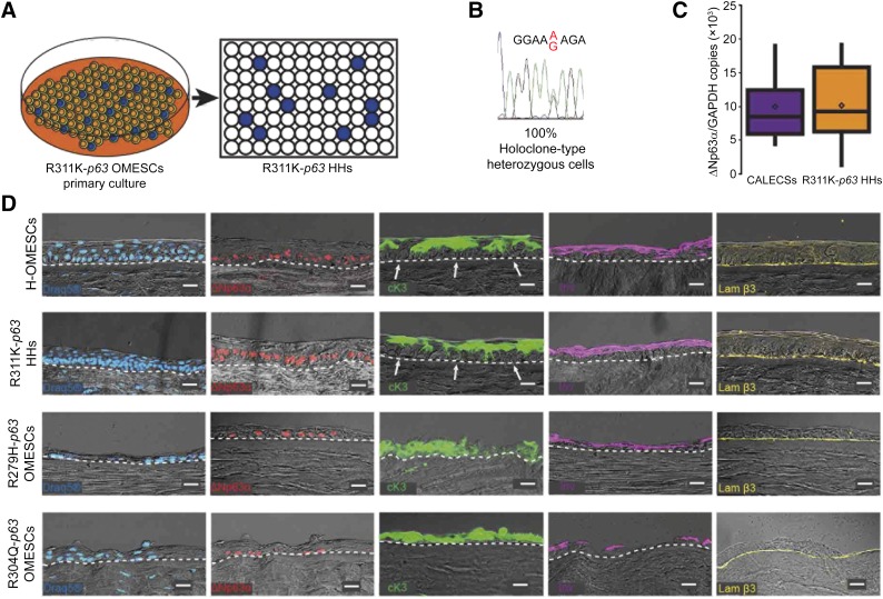
Isolation of R311K-p63 heterozygous holoclones and expression of epithelial cell markers in reconstructed hemicorneas. (A): Cultivation of primary mosaic R311K-p63 OMESCs and isolation of 400 heterozygous R311K-p63 clones through clonal analyses. (B): A representative chromatogram of the sequence around the R311K mutation site of the p63 gene obtained from all the 24 holoclones, previously selected through colony-forming efficiency assay after single-cell clonal amplification. (C): Real-time quantitative analysis of ΔNp63α expression found in CALECSs (n = 19) successfully transplanted in patients with limbal stem cell deficiency, and in R311K-p63 HHs (n = 24). A comparable stem cell content is observed. (D): DRAQ5 staining and expression of epithelial cell markers in reconstructed hemicorneas generated by growing (a) healthy OMESCs, (b) R311K-p63 HHs, (c) R279H-p63 OMESCs, and (d) R304Q-p63 OMESCs onto human keratoplasty lenticules. Cryosections were analyzed through immunofluorescence using ΔNp63α (red), cK3 (green), Inv (violet), and Lam β3 (yellow) antibodies (n = 3) Scale bars = 20 μm. Note that the R311K-p63 HHs’ resulting epithelium was well organized and stratified into four to five cell layers, with basal cuboidal cells differentiating upward to winged cells. The strong expression of the stem cell marker ΔNp63α confirms the maintenance of basal and undifferentiated progenitor cells, which are also negative for cK3 (white arrows). Abbreviations: CALECS, cultured, autologous limbal epithelial cell sheet; cK, cytokeratin; HHs, heterozygous holoclones; Inv, involucrin; Lam β3, laminin β3; OMESCs, oral mucosal epithelial stem cell sheet.

