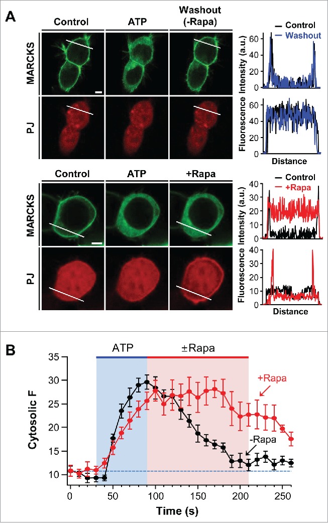
Figure 3.

Depletion of PI4P and PIP2 delays recovery of MARCKS to the plasma membrane. (A) Confocal images and line scanning of cells expressing LDR, PJ, and MARCKS-GFP in response to 50 μM ATP and 1 μM rapamycin. Top and bottom panels represent location change of MARCKS in the absence (black) and presence (blue) of rapamycin, respectively. Bar, 5 μm. (B) Time course of MARCKS translocation in response to the absence or presence of rapamycin. Images of time courses were taken every 5 s by confocal microscope. For analysis, n = 4 .
