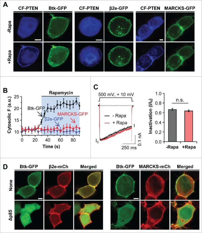
Figure 4.

Effect of PIP3 on subcellular localization of β2e and MARCKS. (A) Confocal images of cells expressing LDR and CF-PTEN with Btk-PH, β2e-GFP, or MARCKS-GFP before and after the addition of 1 μM rapamycin for 2 mins. (B) Time courses of the effects of PIP3 depletion on cytosolic translocation of Btk-PH, β2e-GFP, and MARCKS-GFP. For analysis, n=4. (C) Current inactivation of CaV2.2 channels upon PIP3 depletion. Currents were measured during a 500-ms test pulse to +10 mV. For quantification, n=5. (D) Confocal images of cells expressing Btk-GFP with β2e-mCh or MARCKS-mCh in the absence or presence of dominant-negative p85 (Δp85). Bar, 5 μm.
