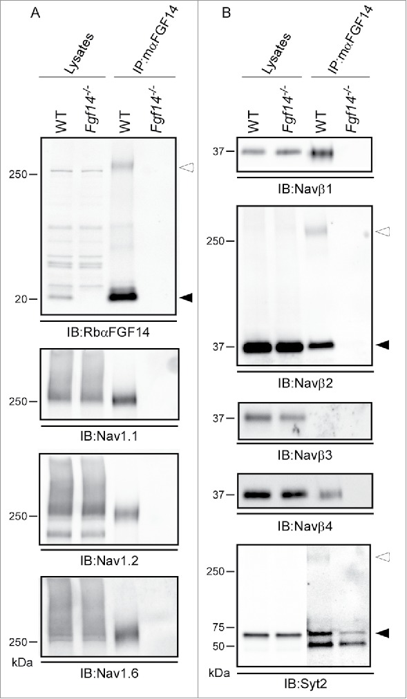
Figure 4.

Western Blot Validation of Selected mαFGF14 Immunoprecipitated Proteins Identified by High Resolution 2D-LC-MS/MS. Representative Western blots of cerebellar lysates from WT and Fgf14−/− mice before and after IP with the mαFGF14 antibody. A. mαFGF14-IP of iFGF14 and Nav α subunits. Immunoblots (IB) with RbαFGF14 revealed a 20 kDa band (closed arrowhead) in the WT lysate and IP, but not the Fgf14−/− lysate or IP. An additional ∼250 kDa band (open arrowhead) is present in the WT IP lane, and likely represents iFGF14 bound to Nav α subunits (see text). IB with the anti-Nav1.1, anti-Nav1.2 and anti-Nav1.6 antibodies revealed that these Nav α subunits are present at comparable levels in WT and Fgf14−/− cerebellar lysates, but only in IP with the mαFGF14 antibody from WT cerebellum. B. IBs with the anti-Navβ1, anti-Navβ2, anti-Navβ3, anti-Navβ4 and anti-Syt2 antibodies revealed comparable levels of each of these proteins in the WT and Fgf14−/− cerebellar lysates. Navβ1, Navβ2 and Navβ4, but not Navβ3, were also identified in Western blots following IP with the mαFGF14 antibody from WT cerebellar lysates. Two bands for Navβ2 were evident in the WT-IPs, a ∼37 kDa band (closed arrowhead) representing Navβ2 and a ∼250 kDa band (open arrowhead), likely corresponding to Navβ2 bound to Nav α subunits. IB with the anti-Syt2 antibody revealed that the ∼60 kDa Syt2 protein (closed arrowhead) was immunoprecipitated with the mαFGF14 antibody from WT and Fgf14−/− cerebellar lysates, but was present at a higher level in the WT IP. The anti-Syt2 antibody also recognized an additional ∼250 kDa band (open arrowhead) in the WT IP, suggestive of Syt2 bound to Nav α subunits.
