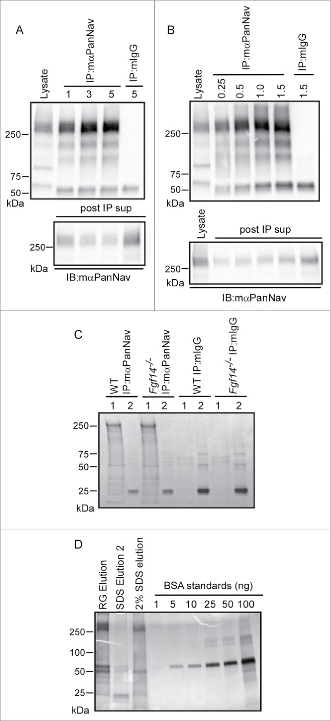
Figure 6.

Optimization of mαPanNav Immunoprecipitations. A. Western blots of WT cerebellar lysates and proteins immunoprecipitated from 0.5 mg of WT cerebellar lysates with variable amounts of the mαPanNav antibody or normal mouse IgG (mIgG). Immunoblotting (IB) with the mαPanNav antibody showed no apparent increase in the amount of Nav α subunit proteins precipitating with 3 or 5 μg (compared with 1 µg) of the mαPanNav antibody. Analysis of the corresponding post IP supernatants (post IP sup) revealed that approximately 70% depletion of the Nav α subunit proteins was achieved with 3 μg of antibody. B. Western blots of WT cerebellar lysate and proteins immunoprecipitated with 3 μg of the mαPanNav antibody or mIgG from variable starting amounts of cerebellar protein. IB with the mαPanNav antibody revealed increasing amounts of Nav α subunit proteins precipitating from samples ranging from 0.25 mg to 1 mg total protein, but no further increase when the starting sample was increased to 1.5 mg protein. Analysis of the corresponding post IP supernatants (post IP sup) revealed that approximately 70% depletion of Nav α subunit proteins from the 1 mg protein sample was achieved. C. SYPRO Ruby stained gel of proteins immunoprecipitated with the mαPanNav antibody or mIgG from WT and Fgf14−/− cerebellar lysates. Beads were eluted first with (1) 2% Rapigest, followed by (2) elution with 1% SDS. Proteins running at the molecular weight corresponding to the Nav α subunits (˜250 kDa) are clearly evident in mαPanNav-IPs from WT and Fgf14−/− cerebella. D. Silver stained gel of proteins immunoprecipitated with the mαPanNav antibody from WT cerebellum. Precipitated proteins were analyzed from beads eluted first with 2% Rapigest followed by 1% SDS elution or beads eluted only with 2% SDS. To estimate the amount of Nav α subunit proteins in each IP sample, a bovine serum albumin (BSA) standard curve was also run. Proteins running at the molecular weight corresponding to the Nav α subunits (˜250 kDa) are clearly evident in the Rapigest and 2% SDS elutions. An estimated 50-100 ng of Nav α subunit proteins are present in the Rapigest elution.
