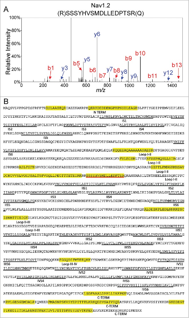
Figure 7.

Mass Spectrometric Identification of Nav1.2 Using In-Solution 2D-LC-MS/MS. A. Representative MS2 fragmentation spectrum of one of the identified Nav1.2 tryptic peptides, corresponding to the sequence NH2-(R)SSSYHVSMDLLEDPTSR(Q) with the y- (in blue) and b- (in red) ions highlighted. B. Amino acid sequence coverage obtained for the mouse Nav1.2 protein following IP of cerebellar Nav channel complexes and LC-MS/MS proteomic analysis. Detected peptides are highlighted in yellow. The peptide for which the fragmentation spectrum is shown (in A) is underlined in red. Transmembrane segments (S1-S6) in each domain (I-IV) are underlined in black. Interdomain cytoplasmic loops I-II, II-III, and III-IV, as well as the N-terminal and carboxyl terminal domains, are also indicated.
