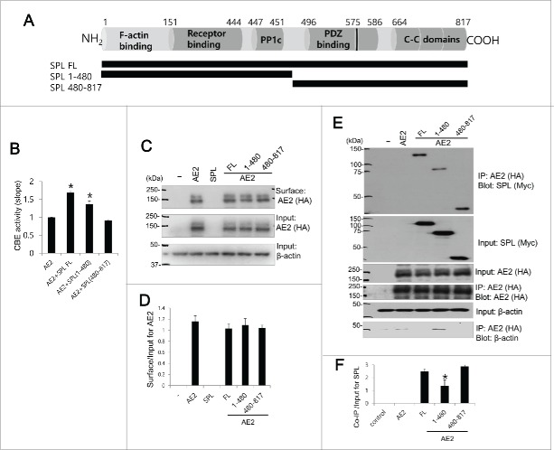
Figure 2.
The essential SPL domain for functional interaction with AE2 Schematic representation of SPL. SPL includes F-acting binding, receptor binding domains, PP1c domain including R-K-I-H-F motif, PDZ binding, another PP1 binding motif within PDZ binding domain (black bar at 575), and 3 coiled-coil (C—C) domains. Schematic diagram of SPL full length (FL), truncated mutants SPL 1–480 and SPL 480–817. HEK293T cells were transfected with AE2 only, or SPL FL, SPL 1–480, and SPL 480–817 with AE2. (B) CBE activity was assessed by the slope of pHi in the absence of Cl− at the beginning of time course (30∼45 sec). The bar graphs show the mean ± SEM. (*P < 0.01). (C and D) The surface expression of indicated plasmids and analysis of surface expression of AE2. AE2 was tagged with HA. Anti-HA antibody was used for the detection of biotinylated proteins. Input actin was used as a loading control. (E) AE2 was tagged with HA and SPL and mutants were tagged with Myc. Anti-HA and anti-Myc antibodies were used for the Co-IP and detection of proteins. Input β-actin was used as a loading control. (F) Analysis of Co-IP of SPL and mutants. The bar graphs show the mean ± SEM. (*P < 0.01). The (-) represented cell lysates.

