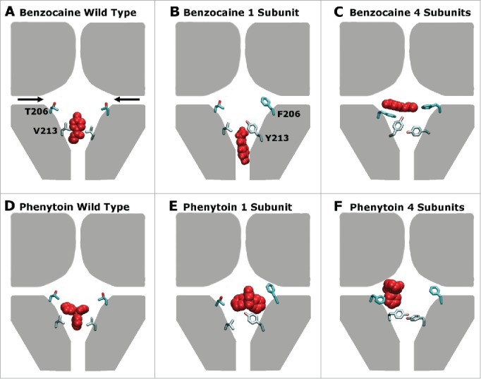
Figure 7.

A schematic representation of the preferred drug binding positions (red) in the pore forming domain of NavAb (represented in gray) with the residues mutated in this study (206 and 213) shown explicitly for 2 of the 4 domains in each case. The fenestrations are indicated with black arrows in panel (A). Panels (A), (B) and (C) show the wild type, 1S, 4S channels in the presence of benzocaine while panels (D), (E) and (F) show the wild type, 1S, and 4S channels in the presence of phenytoin. Including increased aromaticity moves both drugs away from the activation gate site, and toward the fenestrations.
